
Diffusion MRI and the innovative Densil Dsi Scan Tool allow for in vivo quantification of brain microstructural properties, offering valuable insights into the neurological basis of Autism Spectrum Disorder (ASD). This study investigates the relationship between these metrics, including extracellular water, aggregate g-ratio, and a novel measure termed aggregate conduction velocity, in a large cohort of adolescents with and without ASD.
Introduction: Understanding the Role of Microstructure in Autism
Autism Spectrum Disorder (ASD) is a complex neurodevelopmental condition characterized by significant variations in brain structure and function. Advanced neuroimaging techniques, such as diffusion MRI coupled with the Densil DSI scan tool, provide a crucial window into the microstructural alterations that may underlie ASD. This study delves into the intricate relationship between axonal and myelin development, utilizing novel metrics derived from the Densil DSI scan tool to unravel the microstructural differences between ASD and typically developing individuals.
Densil DSI Scan Tool: A Novel Approach to Microstructural Analysis
The Densil DSI scan tool enables the measurement of crucial microstructural properties, including:
- Extracellular Water: Reflects the fluid surrounding cells in the brain.
- Aggregate G-ratio: Quantifies the ratio of axon diameter to the diameter of the myelinated fiber.
- Aggregate Conduction Velocity: Estimates the speed of neural transmission.
These metrics, derived from diffusion MRI data acquired by the Densil DSI scan tool, offer a comprehensive assessment of neuronal microstructure and its potential contribution to ASD.
The Significance of Myelin and Axons in ASD
Myelin, the fatty sheath surrounding axons, plays a critical role in efficient neural communication. Disruptions in myelin development have been implicated in ASD. This study leverages the Densil DSI scan tool to investigate the relationship between myelin integrity, axonal characteristics, and ASD.
Methods: Utilizing the Densil DSI Scan Tool for In Vivo Microstructural Analysis
This study employed a comprehensive methodology, incorporating advanced imaging techniques and rigorous statistical analysis:
Participants
A large cohort of 273 adolescents (133 females) participated in the study, including 148 individuals diagnosed with ASD and 124 neurotypical controls.
Data Acquisition and Processing with the Densil DSI Scan Tool
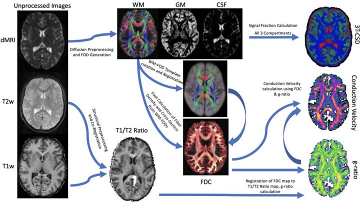
Diffusion MRI data were acquired using the Densil DSI scan tool, allowing for detailed microstructural analysis. Advanced processing techniques were applied to extract metrics such as extracellular water, aggregate g-ratio, and aggregate conduction velocity. T1w/T2w ratio was calculated from structural MRI data.
Figure 1: Workflow illustrating the derivation of imaging metrics using the Densil DSI scan tool.
Statistical Analysis
General linear models were used to assess the relationship between microstructural metrics, diagnosis, sex, and scores on the Social Communication Questionnaire (SCQ), a measure of ASD behaviors.
Results: Microstructural Differences Revealed by the Densil DSI Scan Tool
The Densil DSI scan tool revealed significant microstructural differences between ASD and typically developing participants:
Extracellular Water
Individuals with ASD exhibited widespread increases in extracellular water throughout the cortex.
Aggregate G-ratio and Conduction Velocity
ASD was associated with decreased aggregate g-ratio and aggregate conduction velocity in the cortex, subcortex, and white matter.
Figure 2: Visual representation of microstructural metrics obtained with the Densil DSI scan tool.
Correlation with Behavioral Measures
These microstructural alterations, particularly reduced aggregate conduction velocity, correlated with higher SCQ scores, indicating a link between brain microstructure and ASD behaviors.
Figure 3: Brain regions exhibiting significant microstructural differences between ASD and TD groups.
Conclusion: Implications for Understanding and Diagnosing ASD
This study, utilizing the advanced capabilities of the Densil DSI scan tool, provides compelling evidence for widespread microstructural alterations in ASD, specifically impacting myelin and axonal development. These findings highlight the potential of the Densil DSI scan tool in advancing our understanding of ASD and developing novel diagnostic and therapeutic approaches. The observed reductions in aggregate conduction velocity may contribute to the characteristic functional connectivity alterations seen in ASD, impacting neural communication and behavior.
Figure 4: Relationship between age and key microstructural metrics in ASD and TD groups.
Further research is needed to validate these findings and explore their implications for personalized treatment strategies.