
Understanding the impact of chiropractic spinal manipulation on brain activity is crucial for both musculoskeletal and non-musculoskeletal conditions. This article explores various brain scanning tools, focusing on their spatial and time resolution capabilities, and discusses their potential application in chiropractic research. These tools, including Positron Emission Tomography (PET), Single-Photon Emission Computed Tomography (SPECT), functional Magnetic Resonance Imaging (fMRI), Electroencephalography (EEG), and Magnetoencephalography (MEG), offer unique insights into neuronal activity.
Functional Neuroimaging Modalities: A Comparative Overview
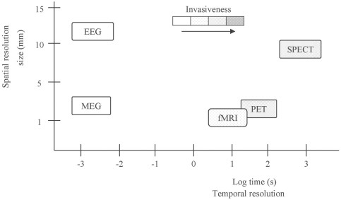
Different functional neuroimaging techniques offer varying degrees of spatial and temporal resolution. Spatial resolution refers to the precision with which a brain scan can pinpoint the location of activity, while time resolution describes how accurately it can track changes in brain activity over time.
Positron Emission Tomography (PET) and Single-Photon Emission Computed Tomography (SPECT)
Both PET and SPECT indirectly measure neuronal activity by detecting changes in regional cerebral blood flow. They utilize radioactive tracers to visualize these changes. PET generally boasts better spatial resolution than SPECT (around 4mm vs 6mm) but both have poor temporal resolution, ranging from minutes per scan. The invasiveness and radiation exposure associated with these techniques limit their use for repeated measurements.
Functional Magnetic Resonance Imaging (fMRI)
fMRI, a non-invasive technique, leverages the blood-oxygen-level-dependent (BOLD) contrast to indirectly measure neuronal activity. It detects changes in blood oxygenation that accompany neural activity. fMRI offers excellent spatial resolution (around 2mm), allowing for precise localization of brain activity. Its temporal resolution (4-5 seconds) is significantly better than PET and SPECT but still slower than electrophysiological methods.
Electroencephalography (EEG) and Magnetoencephalography (MEG)
EEG and MEG directly measure neuronal activity by detecting electrical potentials and magnetic fields generated by active neurons, respectively. EEG offers excellent temporal resolution (milliseconds), enabling real-time monitoring of brain activity. While traditionally considered to have lower spatial resolution (around 10mm), advancements in signal processing techniques are enhancing its localization capabilities. MEG, while more expensive, provides excellent temporal resolution comparable to EEG and good spatial resolution (around 5mm), surpassing EEG in source localization due to minimal distortion from the skull and scalp.
Applications in Chiropractic Research
Functional neuroimaging holds immense potential for advancing chiropractic research. It can help investigate the impact of spinal manipulation on sensorimotor integration, central pain processing, and brain function related to various neurological and psychological conditions. While fMRI offers high spatial resolution for pinpointing affected brain regions, EEG and MEG excel at capturing the temporal dynamics of neuronal responses to spinal manipulation. The choice of technique depends on the specific research question.
Feasibility and Considerations
Cost and access to sophisticated neuroimaging equipment pose challenges to chiropractic research. EEG, with its lower cost and portability, offers a more accessible option for researchers. Technical considerations, such as the need to minimize head movement during scanning, require careful experimental design. Future research should focus on utilizing these powerful tools to explore the neurophysiological mechanisms underlying the effects of chiropractic care. This will not only enhance our understanding of spinal manipulation but also potentially broaden the scope of chiropractic practice.